| CPC G16H 30/20 (2018.01) [G02B 21/365 (2013.01); G06T 3/0006 (2013.01); G06T 5/50 (2013.01); G06T 11/00 (2013.01); A61B 6/5217 (2013.01); A61B 6/5294 (2013.01); G06T 2207/20212 (2013.01)] | 15 Claims |

|
1. An information processing method comprising:
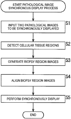
obtaining a first microscopic image and a second microscopic image,
extracting a plurality of tissue areas from the first microscopic image and the second microscopic image using a detection dictionary which is pre-generated through learning using learning cellular tissue region images,
classifying the plurality of tissue areas into groups by image pattern using an automatic clustering technique,
determining a first tissue area of the first microscopic image and a second tissue area of the second microscopic image,
wherein the first microscopic image is at least one of a dark field image, a bright field image or a phase difference image,
wherein the second microscopic image is at least one of the dark field image, the bright field image or the phase difference image,
wherein the first microscopic image and the second microscopic image are images including cell tissue obtained from the same specimen, and
adjusting at least one of shapes, orientations, positions, and sizes of the second tissue area, and causing a display device to synchronously display the first tissue area and the adjusted second tissue area.
|