| CPC G06T 7/11 (2017.01) [G06T 5/30 (2013.01); G06T 7/0012 (2013.01); G06T 7/262 (2017.01); G06V 10/34 (2022.01); G06T 2207/20024 (2013.01); G06T 2207/30104 (2013.01); G06T 2211/404 (2013.01)] | 4 Claims |

|
1. A method for extracting a cardiovascular vessel from a CTA image, comprising the steps of:
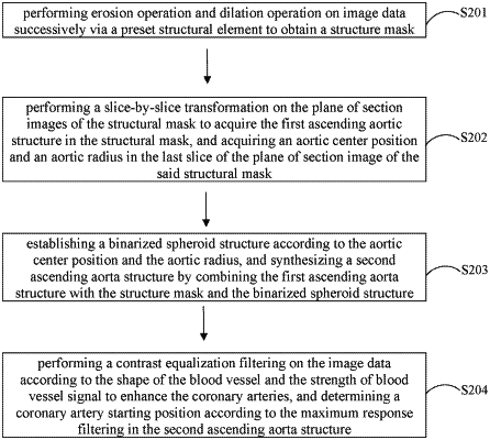
performing a first erosion operation and dilation operation on an image data I successively via a preset structural element Kr to obtain a structure mask I′, said image data I is a coronary angiography image after down-sampling processing targeted at large-size original CTA data; in order to quickly extract a large-size ascending aortic structure without affecting the precision of the structure extraction; and said structural mask I′ is a structure excluding lung region, wherein a sphere of which the radius is controlled at a preset volume element is selected, and the number of the above preset volume elements is 6±2, the sphere of which the radius is controlled at the preset volume element is used as a preset structural element Kr, the erosion operation is performed on the image data I firstly via the preset structural element Kr, and then the dilation operation is performed via the preset structural element Kr to obtain the structure mask I′ with a calculation formula expressed as: I′=I∘Kr=(I⊖Kr)⊕Kr;
performing a slice-by-slice transformation on the plane of section images of the structural mask I′ to acquire the first ascending aortic structure in the structural mask, and acquiring an aortic center position and an aortic radius in the last slice of the plane of section image of said structural mask I′, wherein, during the slice-by-slice transformation, the aortic center positions Co(n) and the aortic radius Ro(n), n=1,2, . . . , N, of the current plane of section image are transformed slice by slice, a preset deviation value is set, and the preset deviation value is ε=6±2, when a distance between the aorta center positions of the current plane of section image and the preceding plane of section image is greater than a preset deviation value, the detection is stopped, and the current plane of section image is determined as the last slice of plane of section image, and the aorta center position Co(n) and the aortic radius Ro(n) in the last slice of plane of section image are acquired;
establishing a binarized sphere structure according to the aortic center position and the aortic radius by taking the aortic radius as the radius of the binarized sphere structure, and synthesizing a second ascending aorta structure by combining the first ascending aorta structure with the structure mask and the binarized sphere structure, wherein at the aortic center position Co(n), a binarized sphere structure Sphx is established by taking the aortic radius Ro(n) as the radius, and a seconding ascending aortic structure AS is synthesized by combining the first ascending aortic structure An with the structural mask B and the binarized sphere structure SphX, and the calculation formula is:
AS=(AN∪SphX)∩B
where AS is the second ascending aortic structure, AN is the first ascending aortic structure, SphX is the binarized sphere structure and B is the structural mask, and erosion is performed via a morphological opening operation, so as to obtain a supplementary area of an ascending aorta root, and the second ascending aortic structure AS is the complete ascending aortic structure of an aortic sinus;
wherein in order to enhance blood vessels of the heart, prevent the contrast of blood vessels in the heart region from being very low and prevent the blood vessel information from being suppressed, a contrast equalization filtering is performed on the image data according to the shape of the blood vessel and the strength of the blood vessel signal to enhance the coronary arteries; FA and FB are blood vessel shape measures, and FC is a blood vessel signal strength measure, which is used to improve the signal-to-noise ratio of blood vessels in the heart region, wherein:
 wherein, RA and RB are two measurement functions based on the characteristics values of Hessian Matrix, RA is used to distinguish between a sheet structure and a linear structure, RB is used to distinguish between a point structure and a linear structure, α, β, and C function as thresholds for controlling the sensitivity of RA, RB, and RC, and γC∈(0,1) is a response expectation constant which generally ranges from 0.5 to 0.8;
under a certain scale σ, spatial Hessian matrix norm ∥Hσ∥=√Σjj≤Dλj2 exhibits a higher response in the lung region with a larger blood vessel contrast, but exhibits a smaller response in the heart region; ∥Hσ∥ mean value and maximum value in the lung region and the peripheral region both trend to monotonously increase;
when Zo0 be a zero matrix, the maximum norm value under each scale is recorded as:
Zσn(x)
 maxx{Zσn−1(x),∥Hσn(x)∥}, n=1, . . . ,N maxx{Zσn−1(x),∥Hσn(x)∥}, n=1, . . . ,N a dynamic threshold c is found to distinguish between the lung region and other tissues according to Zσ≤c and Zσ>c in measurement FC; the non-lung region is defined as θh, the maximum norm is calculated in θh and the full space Θ respectively via rh=∥Hσn(x)∥max/∥Hσn(Θ)∥max, rh∈(0.65,1) is calculated by experimental statistics, and then the parameter c=rh·max(Zσ) is obtained; let RC=c, and a blood vessel characteristic graph V(x, σ) after contrast enhancement is finally obtained,
 where λ2 and λ3 are the second characteristic value and the third characteristic value of the spatial Hessian matrix respectively.
|