| CPC A61B 6/507 (2013.01) [A61B 6/032 (2013.01); A61B 6/463 (2013.01); A61B 6/466 (2013.01); A61B 6/504 (2013.01); G06T 7/0012 (2013.01); G06T 7/12 (2017.01); G06T 7/60 (2013.01); G06T 17/20 (2013.01); G06T 2207/30104 (2013.01)] | 20 Claims |
|
1. A computer-implemented method of determining hemodynamic information for a patient, the method comprising:
receiving medical image data of the patient acquired by a medical image acquisition device, the medical image data including representations of tissue and blood of one or more arterial segments and surrounding area and being characterized through voxels of varying gray scale;
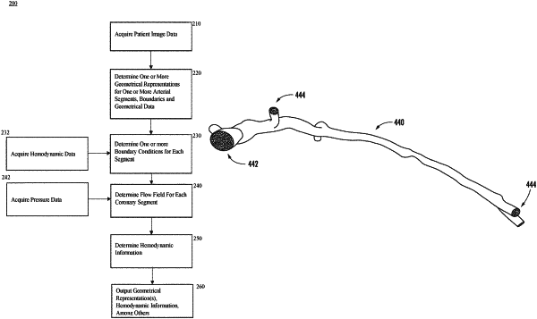
generating a geometrical representation of the one or more arterial segments from the medical image data, wherein a resulting geometrical representation is a three-dimensional model of a spatial volume of the lumens of one or more arterial segments;
determining boundaries and geometry data for each arterial lumen segment, the boundaries of which include a luminal volume and an inflow boundary of the luminal volume for each arterial segment, one or more outflow boundaries, and additional outflow boundaries that each represent a branch or bifurcation in the one or more arterial segments and are disposed between the inflow boundary and a first outflow boundary of the one or more outflow boundaries, the inflow boundary and the one or more outflow boundaries corresponding to a luminal cross-section of the each arterial segment, and the geometry data including a radius for the inflow boundary and a radius for each outflow boundary;
determining boundary conditions for the geometrical representation using the three-dimensional model, the boundary conditions including an interface between the luminal volume and arterial walls, an inflow boundary condition for the inflow boundary based on empirical parameters optimized for determination of a selected hemodynamic parameter of diagnostic interest, and an outflow boundary condition for each outflow boundary, the outflow boundary condition for each outflow boundary being determined using an outflow distribution parameter, the outflow distribution parameter being determined using the geometry data for one or more of the one or more outflow boundaries, the additional outflow boundaries, stored hemodynamic data, or a combination thereof, and a final outflow rate distribution among the outflow boundaries of the segments being determined using a minimization of outflow energy approach encompassing all outflow boundaries;
determining a patient-specific flow field for each arterial segment using a meshing technique within the geometrical representation, and incorporating all of the boundary conditions, the determining including updating outflow distribution boundary conditions as numerical iterations proceed to convergence;
determining hemodynamic information from the flow field, including pressures, velocities and wall shear stresses within the luminal volume and diagnostic hemodynamic indices, including fractional flow reserve, resting pressure ratios, or forces acting on arterial plaques; and
providing an interactive display output of the hemodynamic information that enables receipt of user inputs to manipulate arterial segments, query pressure ratios within the segments, edit vessel segmentation, and assess diagnostic information associated with the hemodynamic information.
|