| CPC G06T 5/50 (2013.01) [G02B 21/367 (2013.01); G06T 5/003 (2013.01); H04N 23/675 (2023.01); H04N 23/959 (2023.01); G06T 2207/10056 (2013.01); G06T 2207/20221 (2013.01); G06T 2207/30024 (2013.01)] | 8 Claims |

|
1. A method for generating a composite digital image of a biological sample from a plurality of images of the biological sample, each of the plurality of images taken along a single axis, the method comprising:
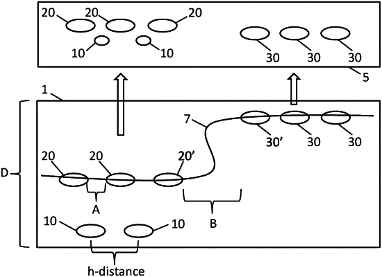
(a) identifying a first focal plane, wherein the first focal plane comprises a first z distance, for a first collection of image objects at a first x-y location of the biological sample, and
(b) identifying a second focal plane, wherein the second focal plane comprises a second z distance, for a second collection of image objects at a second x-y location of the biological sample, and
(c) identifying a first optimal focal plane and a second optimal focal plane based on (a) and (b) respectively, and
(d) combining image objects from a first optimal focal plane and image objects from a second optimal focal plane and generates the composite digital image,
and using a computer apparatus to calculate a sharpness value for the image objects in the first focal plane and calculate a sharpness value for the image objects in the second focal plane and generate a two-dimensional map of the location of the objects having the highest sharpness values, and
identifying an optimal first focal plane and optimal second focal plane for the first and second collection of image objects by performing one or more dilations and erosions on the two-dimensional map of the location of the image objects having highest sharpness values in the first and second focal planes wherein the number of erosions and dilations is proportional to the distance between cell objects of interest in healthy tissue, so as to thereby identify a first and a second optimal focal plane, and wherein image objects located on the first optimal focal plane are presented in-focus and image objects below the first optimal focal plane are deemphasized, and wherein image objects located on the second optimal focal plane are presented in-focus and image objects below the second optimal focal plane are deemphasized, so as to generate a composite digital image of a biological sample.
|