| CPC A61B 8/0883 (2013.01) [A61B 8/12 (2013.01); A61B 8/13 (2013.01); A61B 8/145 (2013.01); A61B 8/483 (2013.01); A61B 8/5207 (2013.01); A61B 8/5215 (2013.01); A61B 8/5261 (2013.01); A61B 90/37 (2016.02); G06T 15/205 (2013.01); A61B 5/055 (2013.01); A61B 8/0891 (2013.01); A61B 8/4245 (2013.01); A61B 8/4254 (2013.01); A61B 8/4483 (2013.01); A61B 8/466 (2013.01); A61B 18/1492 (2013.01); A61B 2018/00357 (2013.01); A61B 2018/00577 (2013.01); A61B 2090/363 (2016.02); A61B 2090/367 (2016.02); A61B 2090/378 (2016.02); A61B 2090/3762 (2016.02); G06T 7/12 (2017.01); G06T 7/344 (2017.01); G06T 13/20 (2013.01); G06T 2200/08 (2013.01); G06T 2207/10132 (2013.01); G06T 2207/10136 (2013.01); G06T 2207/20004 (2013.01); G06T 2207/20081 (2013.01); G06T 2207/20084 (2013.01); G06T 2207/30048 (2013.01)] | 4 Claims |
|
1. A system for generating a three-dimensional segmentation mask, the system comprising:
an ultrasound imager configured to generate two-dimensional intracardiac echocardiography (ICE) images of a part of a cardiac system of a patient with an ICE transducer;
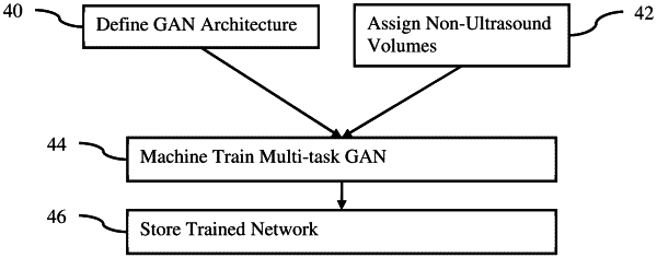
an image processor configured to generate the three-dimensional segmentation mask from the two-dimensional ICE images using a machine-learned multi-task generative adversarial network; and
a display configured to display ablation guidance relative to an image of the three-dimensional segmentation mask.
|