| CPC G02B 21/0012 (2013.01) [G01J 3/0221 (2013.01); G01J 3/28 (2013.01); G01N 21/64 (2013.01); G01N 21/6458 (2013.01); G02B 21/0028 (2013.01); G02B 21/0076 (2013.01); G02B 27/10 (2013.01); G01J 2003/003 (2013.01)] | 17 Claims |

|
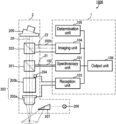
1. An observation auxiliary device for use with a surgical microscope operable to use an exciting light and an observation light as a light source by switching between the exciting light and the observation light, wherein the observation light is a light including a wavelength other than a wavelength of the exciting light, and the surgical microscope includes a first beam splitter and a second beam splitter, wherein the second beam splitter receives light from an observation region observed by the surgical microscope, the observation auxiliary device comprising:
an imaging unit that receives light emitted from the second beam splitter of the surgical microscope and images a plurality of images of the observation region in situations where the exciting light and the observation light are respectively used as the light source;
an output unit that outputs the images imaged by the imaging unit where the exciting light and the observation light are respectively used as the light source;
a spectroscopy unit that splits light emitted from the first beam splitter of the surgical microscope and provides spectroscopy results; and
a reception unit that receives at least one from among an enlargement ratio and a focal length of the surgical microscope;
wherein the output unit corrects at least a portion of the spectroscopy results of the spectroscopy unit according to at least one from among the enlargement ratio and the focal length received by the reception unit to provide corrected spectroscopy results;
wherein the output unit further performs outputting according to the corrected spectroscopy results by the spectroscopy unit;
wherein the spectroscopy unit splits light emitted from the first beam splitter in a situation where normal tissue is observed with the exciting light as the light source and splits light emitted from the first beam splitter in a situation where abnormal tissue is observed with the exciting light as the light source; and
wherein the output unit outputs information relating to a comparison between spectroscopy results for the normal tissue and spectroscopy results for the abnormal tissue, the information output by the output unit comprising a real-time indication that abnormal tissue is included in the observation region.
|